
Intravenous Leiomyomatosis of the Uterus: An Intriguing Case Revealed through Anatomopathological Examination
##plugins.themes.academic_pro.article.sidebar##
##plugins.themes.academic_pro.article.main##
Abstract
Introduction : Intravenous leiomyomatosis (IVL), a rare type of uterine leiomyoma (its incidence is about 0.25% to 0.40% of patients who present uterine fibroma), is characterized by the formation and growth of benign leiomyoma tissue within the vascular wall or lymphatic lumen.
Herein, we presented a case of early stage of IVL successfully treated by surgical removal and a review of actual medical recommendations.
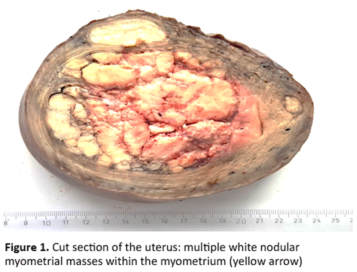
Observation : A 49-year-old woman, gravida 2 para 2, presented to our department with hypogastric pain. On physical examination, a palpable mass in the hypogastrium was noted. Pelvic ultrasound showed a huge uterus with multiple heterogeneous leiomyomas. As the patient was symptomatic and as she had completed their family plan, the decision to perform a total abdominal hysterectomy with bilateral salpingo-oophorectomy was taken. On pathological examination, intravascular growth of benign smooth muscle cell was found within venous channels lined by endothelium. The diagnosis of IVL of the uterus without malignant transformation was confirmed. The patient was monitored for 14 months, and subsequent computed tomography did not reveal any evidence of tumor recurrence.
Conclusion : IVL is a benign, rare and potentially lethal pathology. Clinical manifestations are nonspecific. IVL needs surgical treatment for diagnosis and therapeutic purposes. They require close and prolonged follow-up because of the high risk of recurrence
Keywords:
Benign Neoplasm, hysterectomy, intravenous leiomyomatosis, prognosis##plugins.themes.academic_pro.article.details##

This work is licensed under a Creative Commons Attribution-NonCommercial-NoDerivatives 4.0 International License.
References
- Ziani H, El Idrissi Jallal N, Lahbabi Y, Slaihi Z, Lahbabi S, Oudghiri N, Tachinante R. A case report: intravenous leiomyomatosis extending from the uterus to the right atrium. Ann Med Surg. 2024; 5;86(3):1766-70.
- Wen YL, Ma GT, Miao Q. Diagnosis and treatment of intravenous leiomyomatosis. Zhonghua Wai Ke Za Zhi. 2023; 1; 61(12):1051-7.
- Low HY, Zhao Y, Huang KS, Shen HP, Wu PJ, Tseng CJ. Intravenous leiomyomatosis of the uterus: A clinicopathological analysis of nine cases and literature review. Taiwan J Obstet Gynecol. 2017;56(3):362-5.
- Ma G, Miao Q, Liu X, Zhang C, Liu J, Zheng Y, Shao J, Cheng N, Du S, Hu Z, Ren Z, Sun L. Different surgical strategies of patients with intravenous leiomyomatosis. Medicine (Baltimore). 2016; 95(37):e4902.
- Kdous M, Kraiem NE, Zhioua F, Ferchiou M. Diagnosis and practical management of extra-uterine leiomyoma. Tunis Med. 2015; 93(8-9):582-3.
- Magdalena P, Thomas B, Nina P, Alexander R, Martin A, Christoph N, Josif N, Maja Carina N, Stephan P. Successful one-stage resection of intracardiac intravenous leiomyomatosis: A case report. Gynecol Oncol Rep. 2023; 10;48:101243.
- He J, Chen ZB, Wang SM, Liu MB, Li ZG, Li HY, Zhao G. Intravenous leiomyomatosis with different surgical approaches: Three case reports. World J Clin Cases. 2019; 6;7(3):347-56.
- Zhang G, Yu X, Lang J, Liu B, Zhao D. Analysis of risk factors for post-operative recurrence or progression of intravenous leiomyomatosis. Int J Gynecol Cancer. 2024; 6; 34(5):705-12.
- Barreto-Coelho P, Rosenberg A, Subhawong T, Costa P, Espejo-Freire AP, Bialick S, Jonczak E, Trent JC, D'Amato GZ. Treatment of disseminated intravenous leiomyomatosis with alk targeting crizotinib: a successful case report. JCO Precis Oncol. 2022; 6:e2100336.
- Elbaqqali L, Ait Laayache S, Behraoui H, Zeraidi N, Farhati D, Kharbach A. Intravascular leiomyomatosis of the ute rus. Tunis Med. 2011; 89(12):941-3.